Electronic Bronchoscope
Tuoren Electronic Bronchoscope is used for airway management or for the treatment and diagnosis of the trachea, bronchus, and lungs.
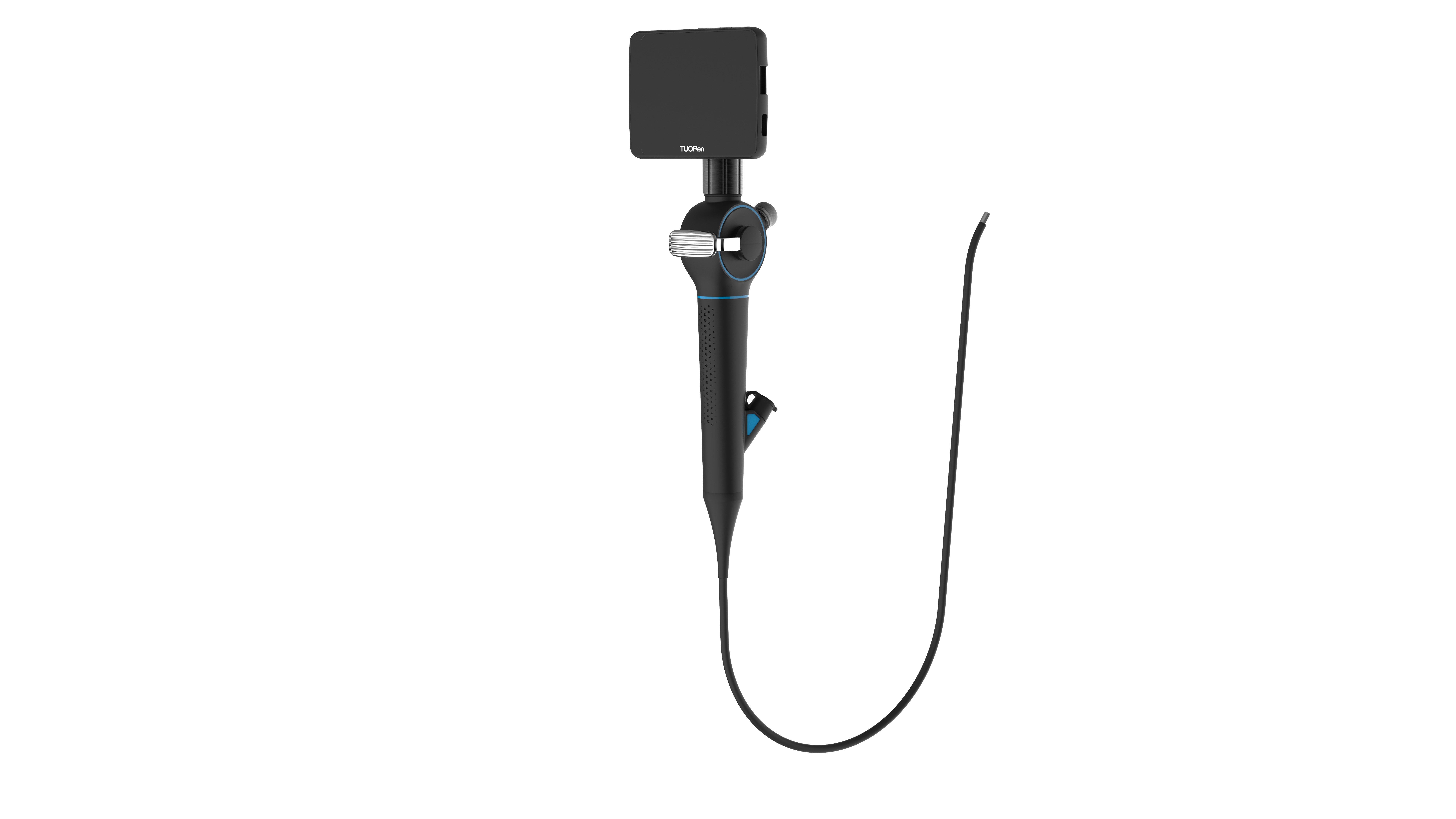
Tuoren offers advanced flexible electronic bronchoscope that no longer applies optical fibre to conduct images, but instead installs a camera on the front end and conducts images with a wire. During the airway management process it is very convenient becauase of its flexible movement. It does not cause any damage to tissues & also helps in minimize irritation to patient.
Features
- Portable: Comes with 3.5″ detachable display, no need for external light source and host.
- Durable: Non-fibre guided, durable, and resistant to bending.
- High-definition display: 3.5″ high-definition display, can be rotated 180° horizontally and vertically, convenient for multi-angle observation and operation.
- Sputum suction and drug delivery: provides a minimum of 1.2mm and a maximum of 2.8mm delivery channel/ working channels to meet the needs of suction and drug delivery.
- Takes photos and videos: can store 1,00,000 pictures or 4 hours of video, provides image data for case analysis, clinical teaching, etc.
- Real-Time View functions on a big screen via an HDMI cable port.
Technical Parameters
|
Specification |
φ3 |
φ7 |
|||
|
Tube Diameter |
2.8mm |
3.8mm |
4.8mm |
5.2mm |
5.8mm |
|
Suction Channel |
No Channel Inside |
1.2mm |
2.2mm |
2.2mm |
2.8mm |
|
Bending Capability |
≥180° |
||||
|
Field of View |
90° |
||||
|
Monitor |
3.5” |
||||
|
Illumination |
≥800 LUX |
||||
|
Operating Hours |
120 Mins |
||||
|
Memory Card |
16GB |
||||
Clinical application
- Inspection and evaluation.
- Treatment.
- Assisted diagnosis and intubation
- Irrigation and Lung Lavage.
Clinical use of Bronchoscope
1. Airway disease diagnosis
- Assessment of difficult airways (such as restricted cervical spine movement, restricted mouth opening, irregular dentition, or vulnerable teeth, etc.)
- Routine airway examination.
2. The treatment of sputum suction, drug administration, biopsy and various treatments, etc.
3. Auxiliary intubation: The bronchoscope can be intubated through the nose and mouth, and the intubation is widely applicable.
- The pre-guided procedure reduces the risk of inability to intubate and ventilate the patient after induction of anesthesia.
- For dual lumen positioning.
- Alignment of bronchial occlusion tube.
- Cerebral intubation with small mouth opening, limited cervical motion, or unstable cervical spine.
- Assess injuries during respiratory contusion or penetrating injuries and avoid the formation of false tracts, and stay away from injuries.
- Intubation is performed to avoid subcutaneous emphysema (subcutaneous emphysema can compress the airway, making it more
- difficult to control).
Accessories Part
- Detachable Monitor – 01
- Baton - 01
- Cleaning/maintenance kit including container r for disinfectant solu1 set- 1set
- Leak Test Kit.
- HDMI Cable.
- Oxygenation Ports- 04
- Working channel Ports- 04
- Brush Biopsy: Short and long.
- Metallic Box to keep the Bronchoscope unit safely.


